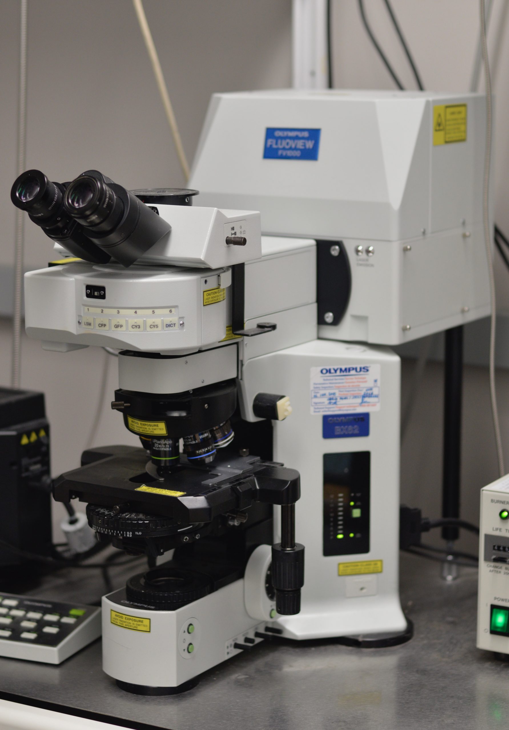
Objectives:
| Name | Magnification | Medium | Numerical Aperture | Working Distance (mm) | Additional Features |
|---|---|---|---|---|---|
| UPLFLN10X2 | 10x | Air | 0.3 | 10 | |
| UMPLFLN10XW | 10x | Water | 0.3 | 3.5 | |
| UPLSAPO20X | 20x | Air | 0.75 | 0.6 | |
| UPLXAPO40X | 40x | Air | 0.95 | 0.18 | Has correction collar |
| LUMFLN60XW | 60x | Water | 1.1 | 1.5 | Has correction collar |
| PlanApoN60xO | 60x | Normal Oil | 1.42 | 0.15 |
Excitation Source:
Diode [405nm], Argon [458nm; 488nm; 515nm], HeNe [543nm; 633nm]
Detectors: PMTs (3 for fluorescence, 1 for transmitted light imaging)
Suggested Software: FV10-ASW ver. 4.02
The NINC Olympus FV1000 confocal microscope saves images in Olympus’ .oib format. Exporting tiffs and jpegs from the FV acquisition software is not recommended as this can cause loss of metadata (e.g. the physical scale of the image). Please download and install the Fiji versions of imagej. Fiji uses the Bio-Formats importer to work directly with .oib files.
Metadata Instrument Identifier: ninc-olympus-fv1000
If you are annotating your data in preparation for data deposit or format conversion (to Neurodata without borders or other file type for long term preservation) please include the above identifier to indicate the data was taken with this instrument.
Serial Number: 435533-9500-000
Citation Guidelines:
If you are publishing your work and have used NINC resources in your project please use the following text including the persistent identifier for NINC (Research Resource ID, RRID):